- Find a Provider
-
Services
-
Redeemer Health provides compassionate care across every stage of life.
- View all Services
- Health Care
- Cancer Care
- Heart Care
- Hospital at Home
- Maternity Care
- Pediatric Urgent Care
- More Health Care Services
-
- Patients & Visitors
- Locations
- Careers
Mammogram
Our mammography sites are right in your neighborhood, offering both convenience and peace of mind.
Breast cancer is the most common cancer among women (except for skin cancer). While a mammogram does not prevent breast cancer, it detects many breast cancers before the effects can be felt. Mammograms are a vitally important screening tool, and we are proud to offer this critical service, throughout Bucks and Montgomery Counties in Bensalem, Feasterville, Huntingdon Valley, Northeast Philadelphia, and Southampton. Don't delay - schedule your mammogram today.
Mammography FAQs
Except for skin cancer, breast cancer is the most common cancer among women. Mammograms do not prevent breast cancer, but they can detect many breast cancers before anyone can feel them. When breast cancer is found in its earliest stages, the majority of patients survive for at least five years.
A mammogram is a low-dose X-ray which produces an image of the breast's inner structures. Mammograms detect tiny calcium deposits or micro-calcifications that are too small to feel. Most of these calcium deposits are benign, but sometimes - especially when in clusters - they may be an early sign of breast cancer.
Traditional Mammography
Mammograms have two types: analog and digital. Analog mammograms record images using film processing, while digital mammograms use X-rays to hit an advanced detector, which send the image data digitally and electronically.
3D Mammography
Breast tomosynthesis (commonly referred to as a 3D mammogram) may be used in conjunction with traditional, digital mammography as part of your annual screening mammogram to capture more breast images.
Unlike a traditional mammogram, a 3D mammogram:
- Uses high-powered computing to convert digital breast images into a stack of very thin layers, or "slices" - building what is essentially a "3D mammogram"
- Has an X-ray arm sweep in a slight arc over the breast, taking multiple breast images in just seconds, producing a 3D image of the breast tissue
3D mammography also offers radiologists the opportunity to view the breast tissue a millimeter at a time, rather than view the complexities of the breast in a flat image.
Automated Breast Ultrasound System (ABUS)
ABUS is done in addition to a traditional or 3D mammogram and is the only breast cancer screening technology that is approved by the FDA for detection in women with dense breast tissue and no abnormal mammography history.
An ABUS helps doctors find cancers hidden in dense breast tissue, which may be missed by traditional screening mammography. Dense breast tissue increases the risk of breast cancer by 4-6 times, making it more difficult to detect cancer in a standard, traditional mammogram.
Unlike traditional or 3D mammography, an ABUS uses sound waves (instead of radiation) to create 3D pictures of the breast tissue. This allows radiologists to look through hundreds of breast tissue images, or "slices." This is different from what is seen with a routine ultrasound, resulting in fewer false negative results.
According to the American Cancer Society, the risk for breast cancer increases with age, especially after age 40. The ACS recommends women ages 40 and older should have a mammogram once per year. Your health care provider, however, may propose another strategy based on your specific health situation. An annual mammogram is what is most common for women ages 40 and older.
The ACS also recommends a clinical breast exam (an examination of the breasts by a health care provider) when a woman has her mammogram.
It's common to be nervous for your first mammogram. Knowing what to expect and how to prepare for your mammogram should help ease your concerns.
Try these tips:
- Schedule your mammogram the week after your period if your breasts are sensitive before and during menstruation.
- Wear a shirt and bra that can be easily removed on the day of your mammogram, as you'll need to undress from the waist up.
- Do not wear deodorant, powder, or lotion. These can affect your X-rays.
- If you have had a mammogram before at a different facility, arrange in advance to have your most recent X-ray sent to us. Your health care provider will want to check for changes since your last mammogram.
For a Traditional Mammogram:
- The technician will help you position your breast on a platform.
- The technician will take two X-rays for each breast, each from a different angle. The machine compresses your breast to get a clear picture of as much tissue as possible, including the armpit.
This process lasts just a few seconds. Having your breast flattened may be uncomfortable, but it shouldn't hurt. If you experience any pain, let the technician know as they can adjust the machine.
For a 3D Mammogram:
3D mammography follows the same two steps above. Your breast will be under compression while the X-ray arm of the machine makes a quick arc over the breast, taking a series of breast images at several angles.
3D mammograms only take a few seconds, and all the images are able to be viewed right there by the technician as they are taken. This allows them to ensure they've captured adequate images to be reviewed by a radiologist.
For an ABUS:
An ABUS takes about 15 minutes. Here's what to expect:
- You'll lie down flat on the exam table.
- Lotion will be applied to your breast.
- A scanner is then firmly positioned on your breast to capture the images.
The radiologist will review your 3D ultrasound images along with your mammogram results.
You may receive your mammography results the same day of your appointment, or the results may be delivered in the mail within a few weeks.
Your doctor will also receive your results. Be sure to follow up with your physician if you haven't been contacted in more than a month. Do not presume that no news is good news - call your physician's office and ask for the results of the report.
Don't panic if your results show an area of concern. Approximately 80 percent of breast lumps are not cancerous. Other tests, such as diagnostic mammograms which take more X-rays, or a biopsy, can help your doctor determine the cause of this result.
Locations
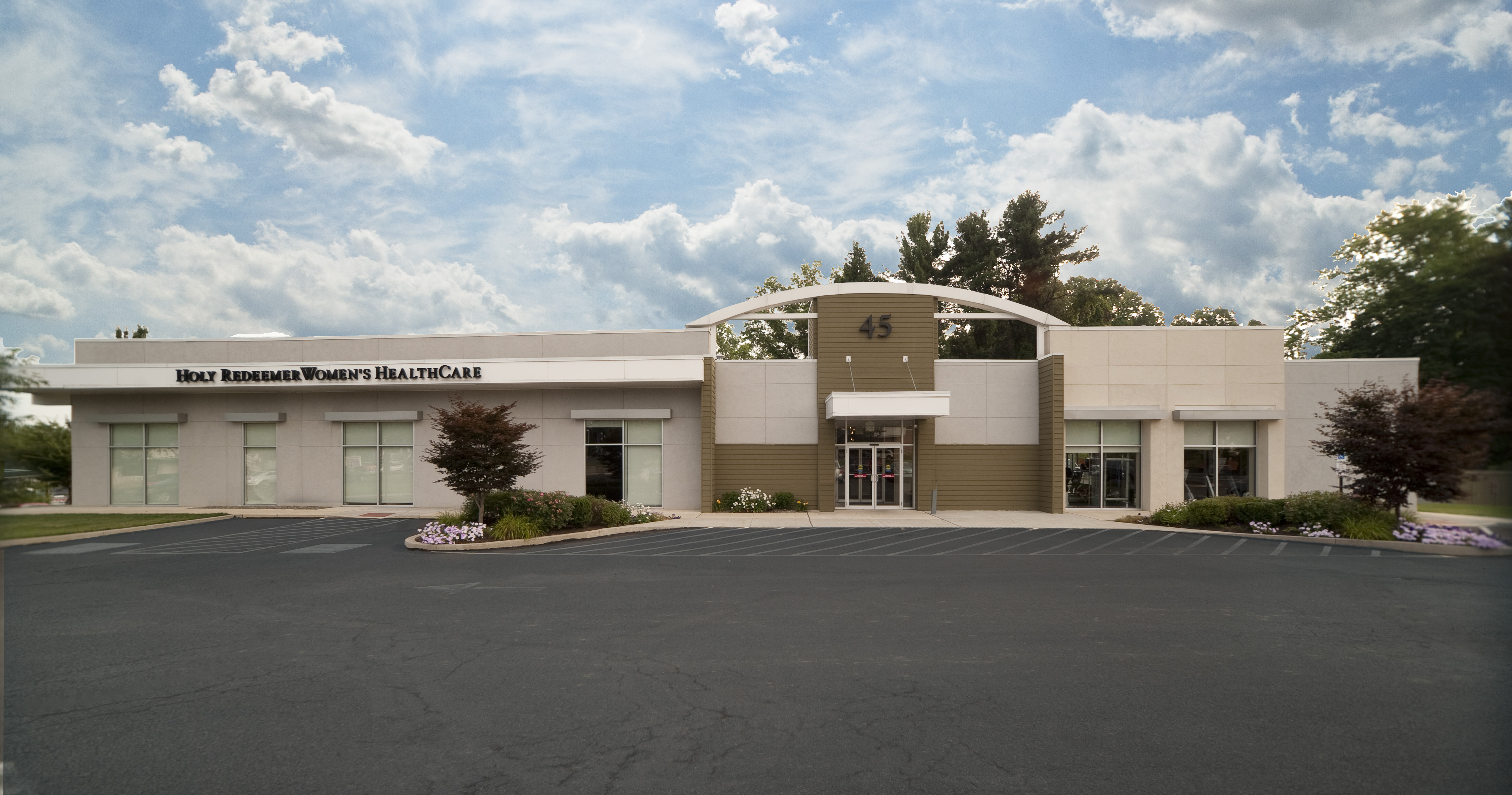
Redeemer Health Mammography - Northeast Philadelphia
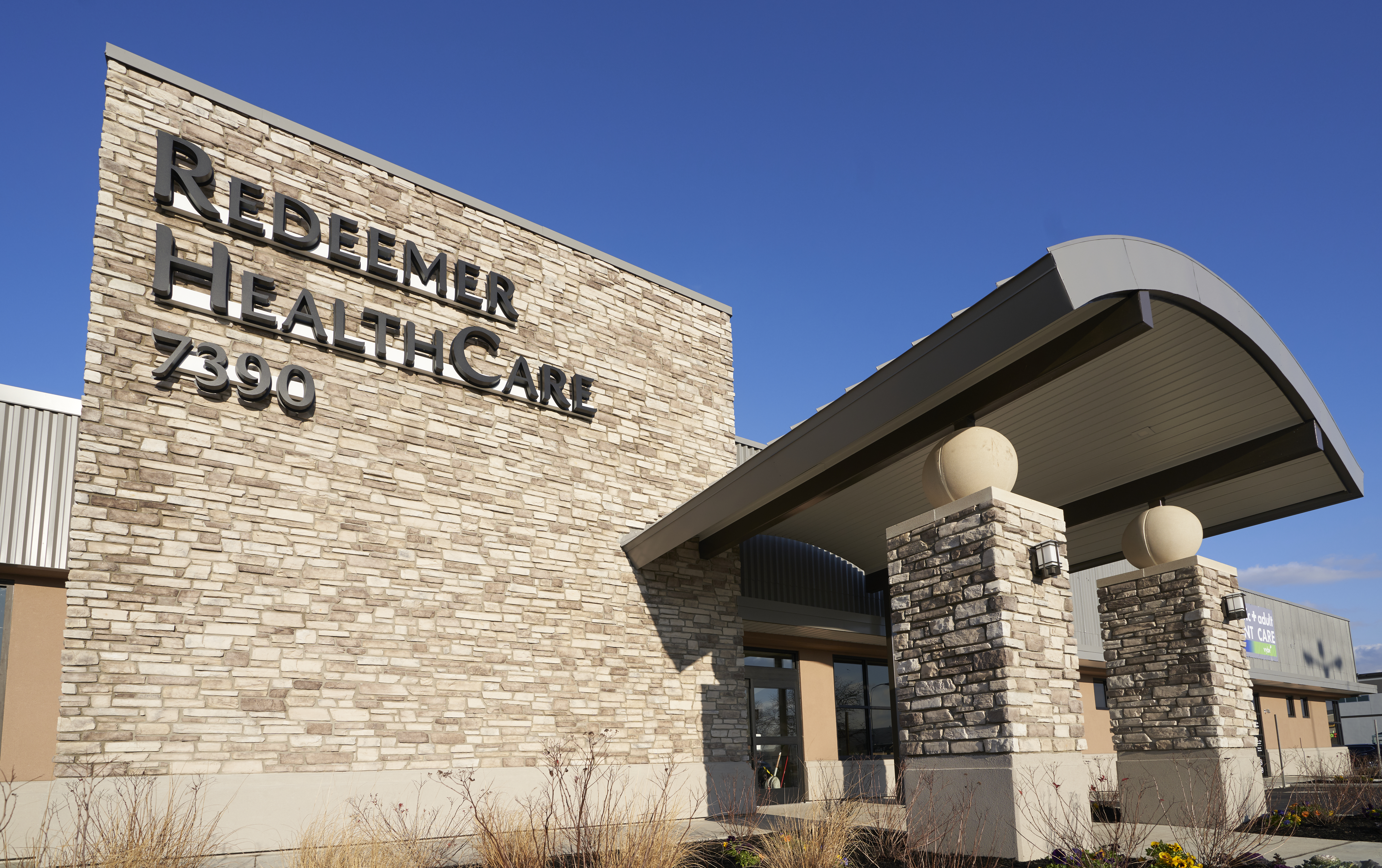
7390 Bustleton Avenue
Philadelphia, PA 19152
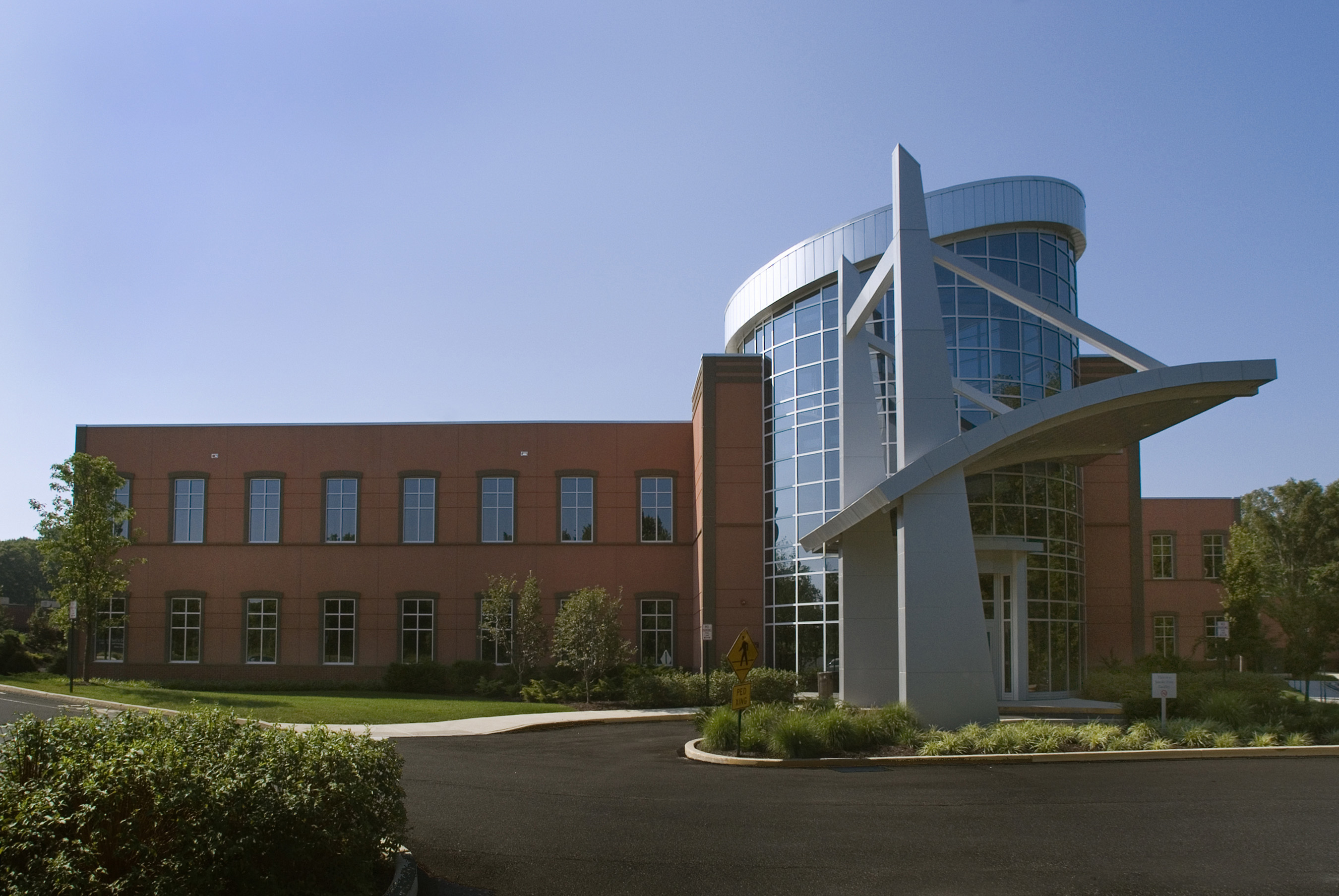
Redeemer Health Mammography - Huntingdon Valley

821 Huntingdon Pike
Huntingdon Valley, PA 19006